© Hanse Equine Hospital

Dr Stephan Leser and Dr Sara Jones perform a vitrectomy (vitreous surgery) on a horse.

Ophthalmology

Ophthalmology is one of the Hanse Clinic’s specialities for horses. Dr Stephan Leser is the senior physician and a recognized specialist in this field. Together with Dr Kirstin Brandt, he performs an average of around 1,000 operations per year both in our clinic and at international partner clinics. Every procedure is minimally invasive and carried out with state-of-the-art equipment.

The minimally invasive procedures help patients recover faster with less pain. Depending on the complexity of the operation and the horse, individual treatments can be carried out under local anaesthesia and without general anaesthesia in a standing position.

In the field of ophthalmology, we offer the following services, among others:

In the field of ophthalmology, we offer the following services, among others:

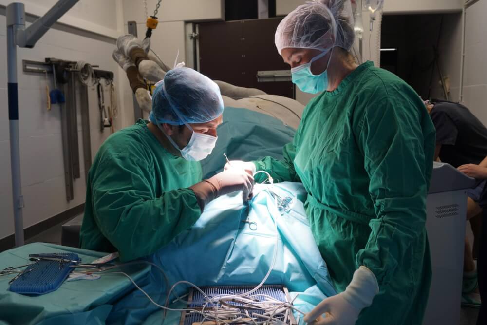
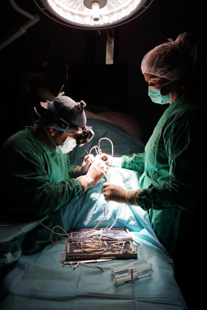
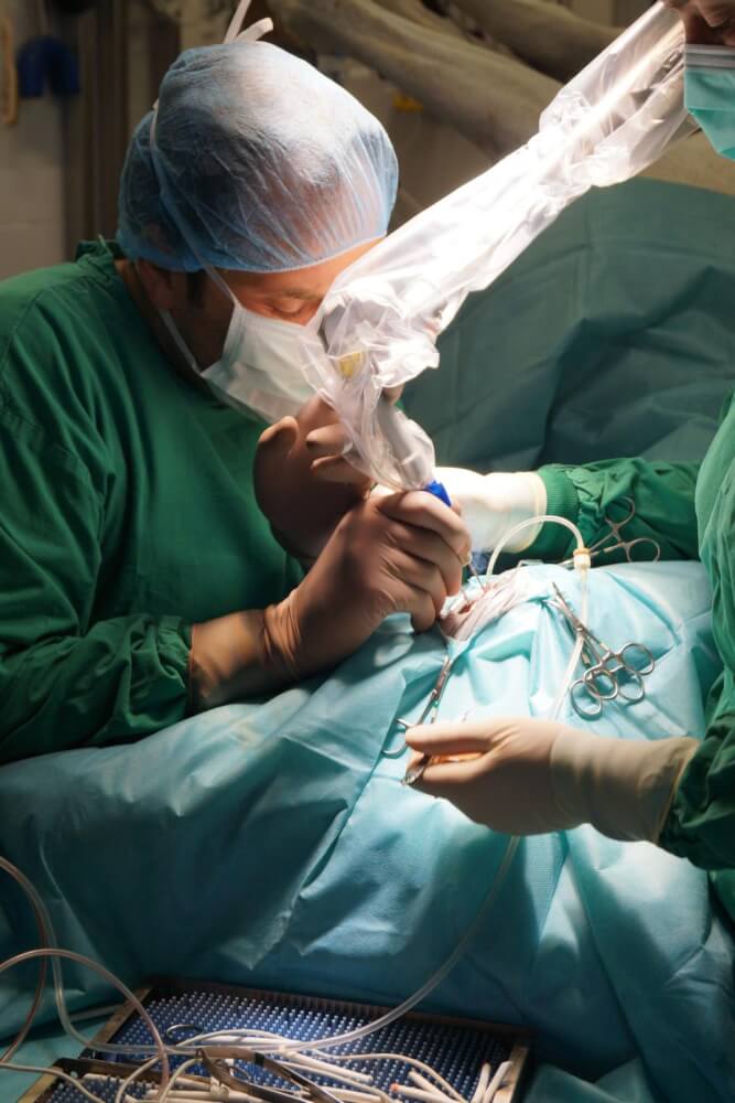
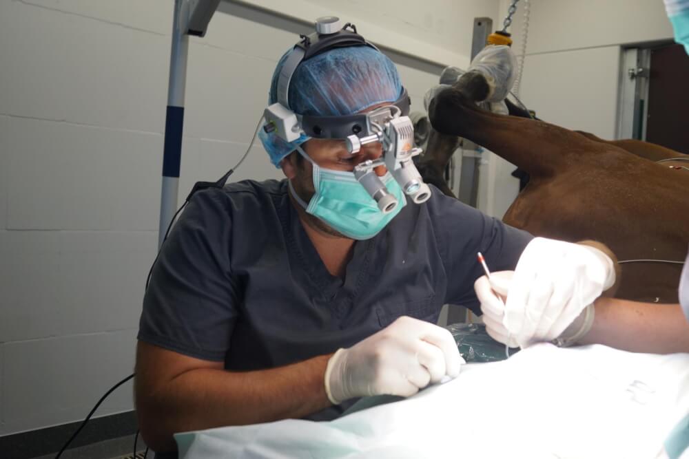
During vitrectomy, the vitreous body is crushed, aspirated and rinsed under sterile conditions. Two different approaches are used for this (double port technique). With the help of a simultaneously cutting and suctioning knife (vitrectome) and an irrigation trocar, inflammatory products and parts of the vitreous body are removed and at the same time the vitreous body space is filled with a balanced salt solution. The accesses are then closed again. The procedure is always performed under general anaesthesia after pre-treatment of the affected eye. The horses remain under in-patient care for a few days after the operation. The most frequent indication for vitrectomy is equine recurrent uveitis, the periodic inflammation of the eye.

The NDR filmed a vitrectomy and summarized it in this report (German):

blank

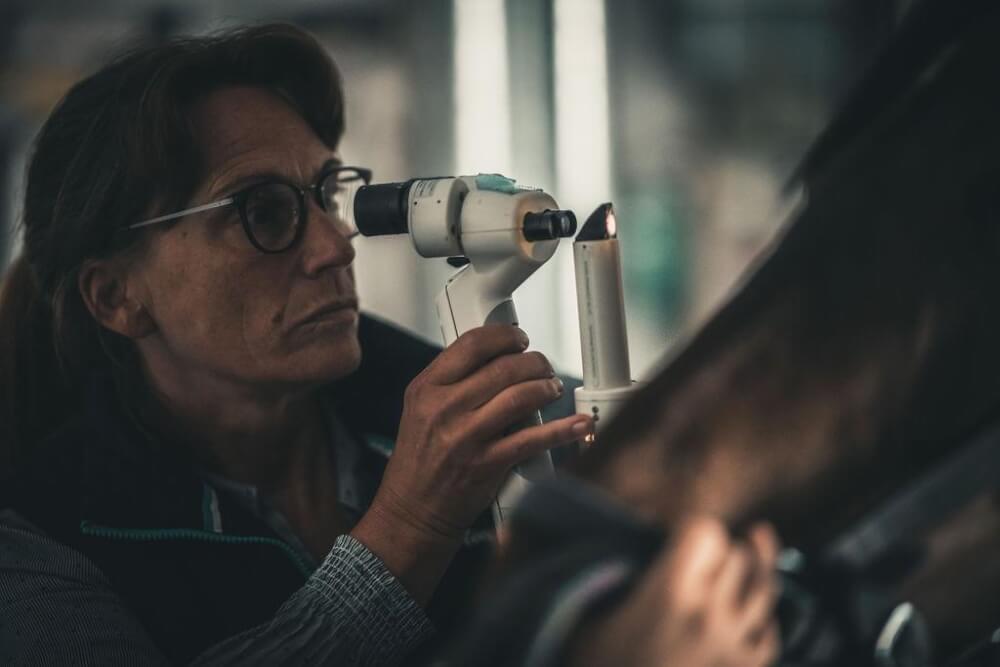
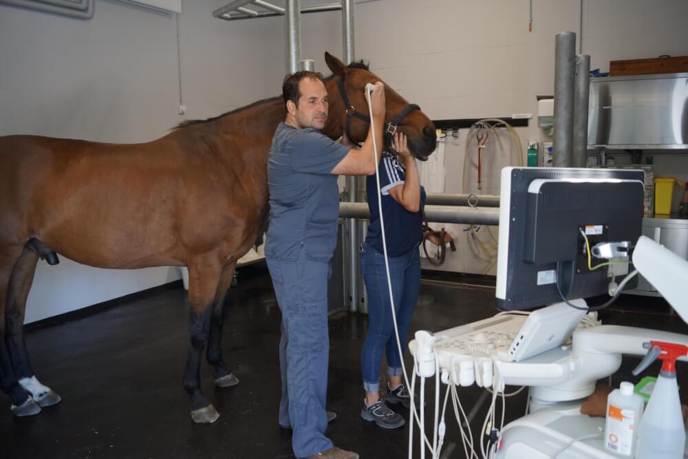
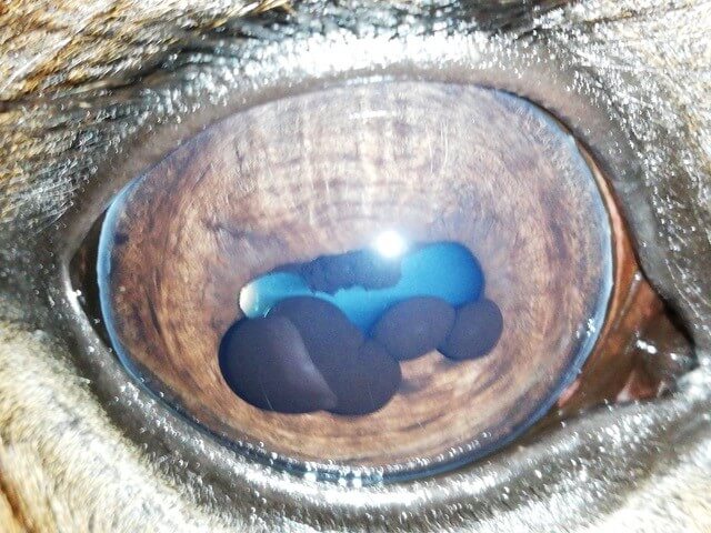
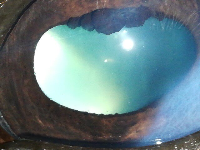
A mature cataract, i.e. the complete clouding of the lens, also known as a cataract, can lead to significant impairment of vision or complete blindness. A cataract can be congenital or acquired. During phacoemulsification, the inside of the lens is crushed and removed using ultrasound waves. Parts of the outer lens capsule remain intact. Prior to this, an eye examination including an ultrasound examination is carried out. Ultrasound examination to check whether phacoemulsification is useful

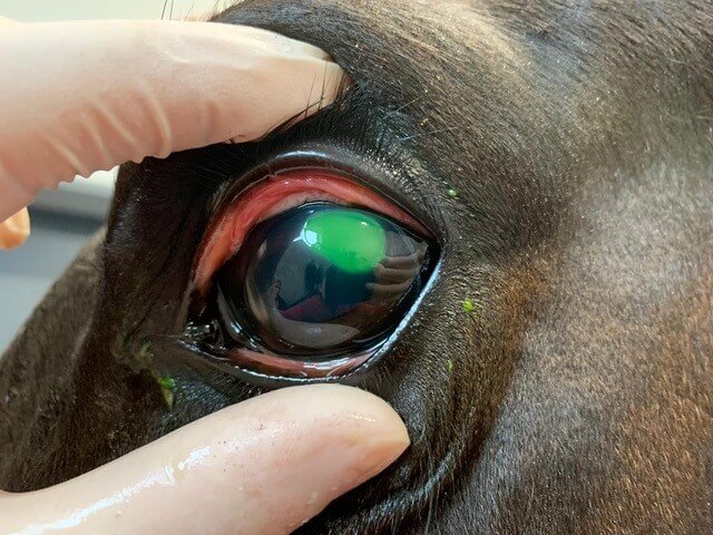
A corneal ulcer can be treated conservatively or surgically, depending on the severity and previous therapy. In the case of deep, extensive and/or infected corneal ulcers, surgical treatment by means of keratectomy (removal of diseased cornea) and conjunctivoplasty is indicated.. For this purpose, a part of the conjunctiva (conjunctiva) is prepared and sewn into the cornea. The diseased area is thus stabilised and can heal faster due to the vessels contained in the conjunctiva. Horses require intensive local aftercare for some time and are often fitted with a subpalpebral catheter (eyelid catheter) to minimise manipulation of the eye and allow optimal application
A corneal injury is usually caused by trauma and can pose a serious threat to vision as well as to the eyeball. A very deep injury is an absolute emergency and requires the fastest possible treatment. Depending on which structures of the eye are involved, surgical treatment can be performed by a corneal suture with or without conjunctivoplasty..

The cornea is exposed to a variety of pathogens every day. If the barrier function of the tear film and cornea is disturbed by injuries or immunological processes, a corneal infection by bacteria or fungi can occur. Depending on the severity, intensive treatment with medication or surgery may be necessary. Deep corneal infections often lead to tissue destruction and the development of an ulcer. The infected tissue must be thoroughly removed and the affected cornea is usually treated with conjunctivoplasty. Fungal corneal infections (keratomycoses) in particular are on the increase and are often difficult to treat. Intervening as early as possible is important for the best possible prognosis.

Glaucoma is a painful disease and rarely occurs in horses as a late consequence of periodic ocular inflammation (equine recurrent uveitis, ERU), after trauma or tumours in the eye Glaucoma is characterised by a pathological increase in intraocular pressure. As a rule, the aqueous humour produced by the ciliary body can only partially drain away and too slowly. This causes symptoms such as corneal opacity (oedema), enlargement of the eyeball and eventually blindness. Laser cyclophotocoagulation reduces aqueous humour production by using laser energy to partially obliterate the ciliary body inside the eye. This can lead to a reduction in intraocular pressure. The procedure can be performed on a standing, sedated horse under local or general anaesthesia.

Inflammation of the cornea, keratitis, is often a recurring clinical picture and in some patients can only be insufficiently controlled by local therapy. During keratectomy, the removal of chronically diseased corneal tissue, part of the cornea is lifted off and removed with special instruments. In this way, local antigens that can always lead to inflammation can be removed. Depending on the severity and location of the keratitis, a fairly large wound area can develop, which can take several weeks to heal. If there are no complications, the eye is free of irritation and the patient has good vision.

Paracentesis involves puncturing the anterior chamber of the eye using a very fine cannula. A small amount of aqueous humour can be removed and analysed in the laboratory. Here, the focus is primarily on the examination for leptospires to clarify a leptospire-associated internal ocular inflammation (equine recurrent uveitis, ERU).. Paracentesis can also be used to place small amounts of various medications in the anterior chamber of the eye. Paracentesis is possible under general anaesthesia as well as on the standing-sedated horse under local anaesthesia.

Almost 10% of all horses worldwide suffer from the so-called equine recurrent uveitis or colloquially also called periodic eye inflammation. Equine recurrent uveitis (ERU) is a disease that can lead to progressive damage up to blindness and even loss of the eye due to recurrent inflammation of the inner eye.

With each bout of inflammation, the structures inside the eye are further injured and the associated pain increases rapidly. If ERU is not noticed and treated in time, it almost always leads to blindness of the horse and in the worst case the affected eye has to be surgically removed.

ERU can have various causes. However, research has shown that the so-called leptospires – bacteria that can be found in every stable – are mainly responsible for it. Leptospires enter stagnant water, feed or bedding of horses especially through rodent urine. If these bacteria get into the horse’s body, they are normally fought off by the immune system and pose no threat to health.

However, if the immune system cannot render these bacteria harmless, it sometimes happens that the bacteria reach the inside of the eye and settle in the vitreous body. The bacteria infect the vitreous body and inflammatory products are formed that can no longer be removed from there. In 25 – 30 % of all horses suffering from ERU, both eyes are affected. ERU is not transmissible from horse to horse and therefore poses no danger to neighbouring horses.

If the inflammation occurs in the posterior segment of the eye, the symptoms are hardly visible to the owner. After diagnosis, many owners report long-lasting performance deficits, nervousness and a change in character. If the inflammation is in the anterior structures of the eye, the pain is clearly visible to the owner. The horses show eyelid pinching, have increased lacrimation and are sensitive to light. Closer examination of the affected eye often reveals a clouded cornea and a constricted pupil.

Recurrent episodes of inflammation cause more and more damage to the inner structures of the affected eye. The lens becomes increasingly cloudy due to more and more adhering inflammation products and adhesions of the iris to the front surface of the lens. This can lead to a cataract. The intraocular pressure drops and the eyeball shrinks. In the final stage, complete detachment of the retina occurs, leading to blindness of the diseased horse.

Prof. H. Gerhards introduced vitrectomy as an effective method for treating ERU in 1991. In vitrectomy, the infected vitreous body, including bacteria, inflammation products and opacities, is removed from the vitreous cavity through an intraocular operation. The vitreous cavity is simultaneously filled with a fluid similar to aqueous humour without any loss of pressure.

This treatment is carried out at the Hanse Equine Hospital more than 1000 times a year and is an absolute routine for the Head of Ophthalmology, Dr Stephan Leser and his team.

A horse suffering from ERU cannot recover on its own. Thanks to the minimally invasive vitrectomy, however, more than 97% of the horses are free of the disease. The horse can see again immediately afterwards and no longer suffers any pain.

Various tumours can occur in the area of the eyelids, the conjunctiva or on and in the eye itself. Using a CO2 laser to remove tumours is a low-bleed method and can reduce the risk of recurrence by obliterating the wound bed. When circumferential increases are noticed, quick action often improves the prognosis. Tumour resection by laser can also be combined with other local therapies, depending on the case.

A posterior synechia, a pathological connection between the iris and the anterior surface of the lens, usually develops in acute uveitis. Diese geht häufig mit einem starken Pupillenkrampf einher, der zusammen mit der inneren Augenentzündung zum Anhaften der Iris an der Linsenkapsel und einer dauerhaften Verbindung führt. Posterior synechiae can cause the pupil to become non-circular and impaired in function. This can lead to a reduction in visual acuity. For synechiolysis, the anterior chamber of the eye is punctured and the synechiae injected with hyaluronic acid. Very fine instruments are then used to lift the iris and detach it from the lens capsule.

Eyelid injuries are common in horses and should always be treated by a veterinarian, because adequate eyelid closure is necessary for undisturbed corneal function. Eyelid injuries can often be treated on the standing-sedated horse. Eine genaue Untersuchung der umliegenden Strukturen ist vor allem bei Traumata wichtig und kann auch eine röntgenologische und ultrasonografische Untersuchung umfassen.

Various conditions, such as tumours on the eyelids or extensive injuries with tissue loss, can lead to loss of the eyelid margin and prevent adequate eyelid closure. Since complete eyelid closure is essential for the function of the eye, a gradual restoration must take place. Reconstruction of the eyelid margin is implemented by various surgical techniques, depending on the case, and may include displacement plasty, skin graft techniques or laser surgery.

The removal of the eye, bulbous extirpation, may be indicated for painful, blind eyes or severe diseases of the inner eye, such as tumours, among others. In most cases, the procedure can be performed on a standing sedated horse under local and conduction anaesthesia After the eye is removed, the eyelid margins are adapted and the skin gradually sinks into the eye socket. The horses first receive a head bandage and can be discharged after a short stay in the clinic.

Diseases of the anterior chamber of the eye are commonly seen in horses and can have various causes, such as traumatic, inflammatory or tumorous. In case of inflammation or infection, injections can be made into the anterior chamber of the eye (intracameral injection). In most cases, these can be performed on the standing-sedated patient.
In most cases, these can be performed on the standing-sedated patient. Often they are not a problem for the horses, but above a certain size they can lead to visual impairment as well as irritation of the innermost layer of the cornea (endothelium). In these cases, they can be reduced in size and sclerosed by means of puncture and the use of laser energy.
More extensive operations in the anterior chamber of the eye are performed under general anaesthesia. In this procedure, various instruments are inserted into the anterior chamber of the eye via a tunnel-shaped, limbal access (transition from the choroid to the cornea). To protect the eye from collapsing and damaging the back of the eye, part of the aqueous humour is aspirated and replaced with hyaluronic acid. This technique can also be used to remove larger tumours, such as iris melanomas. More rarely, foreign bodies occur in the anterior chamber of the eye, for example due to trauma. They can also be removed via a tunneled limbal approach (transition from choroid to cornea) with the help of instruments.

Electroretinography is used to check the functionality of the retina. This is why it is always indicated when the cause of a visual impairment is suspected to be changes in the area of the retina or when the fundus of the eye cannot be adequately assessed due to opacities in the inner eye. For example, an electroretinogram (ERG) may be useful in cases of equine recurrent uveitis (ERU), retinopathies, congenital stationary night blindness (CSNB) or in differentiating between peripheral and central blindness.
To create an ERG, the retina is stimulated with flashes of light.
This creates electrical potentials that can be derived, measured and displayed in a characteristic curve.
The procedure is performed on a standing-sedated horse.